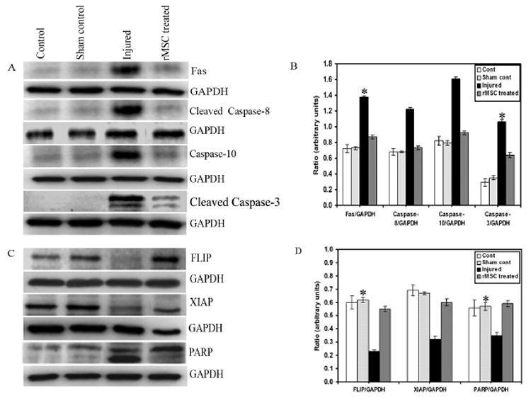
Fig. 7. Immunoblot analysis of caspase-3-mediated apoptotic pathway in spinal cord sections.

Equal amounts of protein (40 μg) were loaded onto 10%-14% gels and transferred onto nylon membranes, which were then probed with respective antibodies. The blots were stripped and reprobed with GAPDH to assess protein levels. A and C shows the Fas and caspase-3 mediated apoptotic pathway proteins with respect to GAPDH; B and D are their quantitative estimations, respectively. Inhibition of the caspase-3-mediated apoptotic pathway by rMSC shows downregulation of Fas and caspases, upregulation of FLIP, XIAP, and inhibition of PARP cleavage. Antisera for caspase-8 and caspase-3 recognize cleaved fragments also. Each blot is representative of experiments performed in duplicate with each sample (n = 3). Error bars indicate SEM. * Significant at p <0.05.
