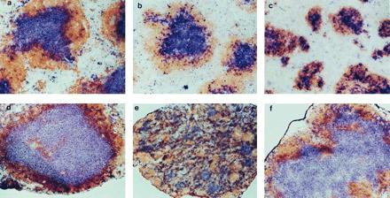
Figure 1.

Disturbed follicular segregation of T cell and B cell zones in LNs of TNFR-I−/− mice and spleen of LT-α−/− mice. Wild-type (a and d), TNFR-I−/− (b and e), and LT-α−/− (c and f) mice were immunized i.p. with 108 SRBCs, and then 10 days later spleen (a–c) and LNs (d–f) were harvested and sections were prepared and stained with anti-Thy1.2 antibody (blue) and anti-B220 (brown) to visualize the T cell and B cell zones, respectively. (a–d and f, ×100; e, ×40.)
