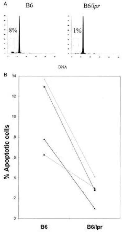
Figure 4.

(A) Spleen cells stained with anti-CD4 from B6 (Left) and lpr mice (Right) were cultured overnight after irradiation and assessed for apoptosis by quantitating DNA content. Data shown represent the DNA content of CD4+ cells from a 7-month-old animal and are from a representative experiment. (B) The percent apoptosis of CD4+ B6 (Left) and lpr (Right) irradiated spleen cells that were cultured as in Fig. 1 is shown. Data points represent individual experiments where three spleens were pooled in each group per experiment. Data from the same experiment are connected with a line. The mean % apoptotic CD4+ T cells from B6 was 19.1 ± 6.3 and 5.4 ± 1.8 for B6/lpr. Numbers after the mean represent the standard error. The difference between the groups was significant to a P value of 0.0383.
