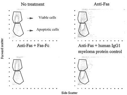
Figure 9.

Thymocytes from three pooled 3- to 4-month-old B6 mice were cultured overnight (Upper Left), incubated with 1 μg/ml of anti-Fas during culture (Upper Right), incubated with 1 μg/ml of anti-Fas with the addition of 100 μg/ml of Fas-Fc during culture (Lower Left), or incubated with 1 μg/ml of anti-Fas with the addition of 100 μg/ml of a purified human IgG1 myeloma protein during culture (Lower Right). Viable and apoptotic cells are depicted in the outlined regions as labeled. Viable and apoptotic cells in each panel are as follows: no treatment, 63% viable and 21% apoptotic; anti-Fas treatment, 24% viable and 44% apoptotic; anti-Fas plus Fas–Fc, 57% viable and 22% apoptotic; and anti-Fas plus human IgG1 myeloma protein control, 22% viable and 51% apoptotic.
