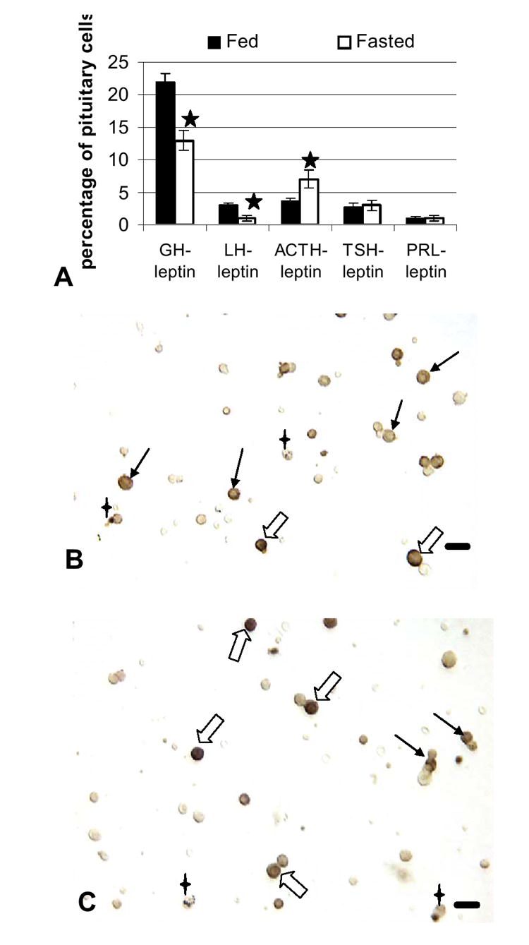
Figure 4.

Pituitary cells that express leptin: Effects of food deprivation Dual labeling for leptin proteins and each of the pituitary hormones was done on freshly dispersed pituitary cells. Figure 4A shows a significant decrease in percentages of AP cells with leptin proteins and GH or LH and an increase in percentages of cells with leptin proteins and ACTH. Stars=values different from those of fed rat cell populations. Figure 4B illustrates a field dual labeled for leptin (black) and ACTH (orange) proteins. Black, filled arrows show corticotropes labeled for only ACTH and the 4 pointed stars indicate cells labeled for only leptin. Dual-labeled corticotropes are indicated by the hollow arrows. Figure 4C shows a field from fasted animals, similarly labeled for leptin and ACTH. The dual labeled cells (hollow arrows) are more darkly labeled for leptin, making the ACTH label difficult to see in a lower magnification image. Cells that express only leptin (4 point star) or ACTH (black arrows) remain in the populations from both groups. Bar=15 μm.
