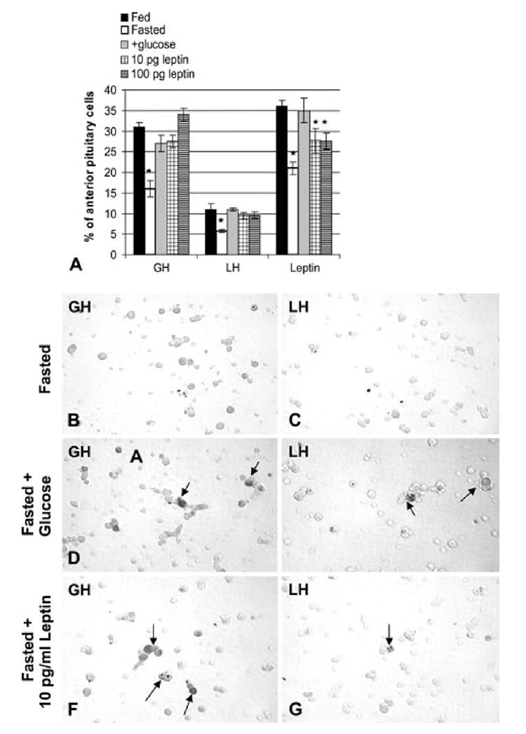
Figure 7. Restorative effects of glucose, in vivo, or leptin, in vitro, on GH, LH and leptin expression.

Counts of cells with leptin, GH, and LH proteins labeled by immunocytochemistry. In Figure 7A, these counts represent the latest groups of fasted animals, including those receiving glucose. Note that the decline in cells with leptin proteins is greater in this group of fasted rats, than in the first group shown in Figure 3A. All three gene products are recovered in the fasted rats receiving glucose (+glucose). Figure 7A also illustrates the restoration of GH and LH cells in cells from fasted rats, that received 10 pg/ml or 100 pg/ml leptin for 1 h, in vitro. Cells bearing leptin are partially restored in the presence of leptin. Figures 7B and C illustrate pituitary cells from fasted rats immunolabeled for GH (Fig 7B) or LH (Fig 7C). Figures 7D and 7E illustrate fields from fasted rats receiving glucose water immunolabeled for GH (Fig 7D) or LH (Fig 7E) showing the recovery in GH or LH cells. A similar recovery, in vitro, is illustrated in Figures 7F (GH cells) or 7G(LH cells). These cultures were from fasted rats receiving 10 pg/ml leptin for 1 h, in vitro. The vehicle control for this group is identical to the cells depicted in Figures 7B and 7C, having few GH or LH cells after 1 h in culture. This 1 h treatment, in vitro, with exogenous leptin has the same restorative effect on GH and LH cells as glucose, in vivo. Bar=21 micrometers.
