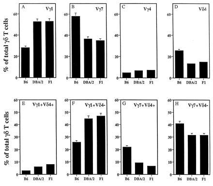
Figure 1.

Representation of different γδ i-IEL subsets in B6, DBA/2, and B6D2F1 mice. (Upper) i-IEL from the three mouse strains were isolated and stained with fluorescein isothiocyanate-labeled anti-pan γδ mAb and biotin-labeled anti-Vγ1 (A), anti-Vγ7 (B), anti-Vγ4 (C), or anti-Vδ4 (D) mAbs followed by streptavidin–phycoerythrin and analyzed on a FACScan. Data are shown as the percentage of total γδ i-IEL expressing the indicated V region and correspond to the mean ± SD of six independent determinations using two to four animals per determination. (Lower) The same i-IEL preparations were stained with fluorescein isothiocyanate-labeled anti-Vγ1 (E and F) or anti-Vγ7 (G and H) mAbs together with biotin-labeled anti-Vδ4 mAb followed by streptavidin–phycoerythrin and analyzed as above.
