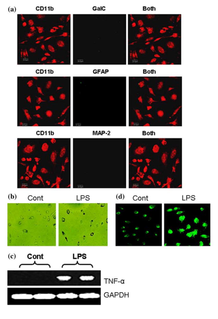
Fig. 3.

To examine the purity of microglia, cells were double-immunolabeled with CD11b and either GalC, GFAP or MAP-2 and observed under a confocal laser-scanning microscope (a). Microglial cells were either stimulated with LPS (1 μg/ml) in serum free condition or treated with serum free media alone and after 3 h cells were exposed to latex beads for 90 min at 37°C to monitor phagocytosis (b). Microglia were treated with LPS under serum-free condition. After 6 h of treatment, total RNA was analyzed for the expression of TNF-α by semi-quantitative RT-PCR as described under “Materials and Methods” (c). Microglia were treated with LPS and after 24 h of treatment, cells were immunostained with antibodies against CD11b (d). Results are representative of three separate experiments
