
Figure 2.
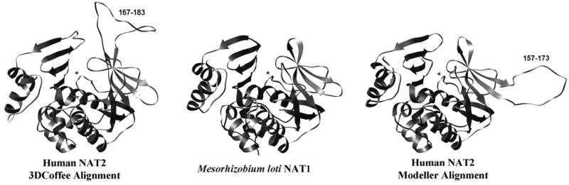
Homology models of human NAT2, and the Mesorhizobium loti NAT1 bacterial template structure (shown for reference). The Modeller alignment produces a second domain loop away from the active site, whereas the 3DCoffee alignment produces a second domain loop adjacent to the active site pocket. Asterisks identify the active site loop, and the residue numbers of each insert are shown.