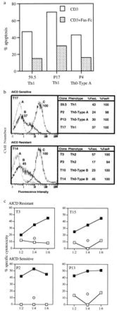
Figure 4.

(a) AICD is inhibited by Fas–Fc fusion protein. Apoptosis-sensitive clones 59.5, P4, and T17 were stimulated with anti-CD3 antibodies in the absence (□) or presence (▪) of 10 μg/ml of Fas–Fc fusion protein and assayed for apoptosis by ELISA. Fusion protein Fas–Fc is composed of human Fas and a human immunoglobulin constant region. (b) Expression of FasR and FasL on subsets of T cell clones. For immunofluorecence, 1 × 10⁶ T clones (59.5, T17, P3, T3, P2, P13, T10, and T14) were stimulated with anti-CD3 antibodies in the presence (for FasL) or absence (for FasR) of 25 μM of the metalloprotease inhibitor KB8301. Addition of KB8301 prevents FasL cleavage, resulting in high levels of cell surface expression of FasL After 3 hr the cells were washed in PBS containing 2% serum and reacted with biotinylated antibody to FasR (C) or to the FasL (B) (PharMingen) or no antibody (A) for 45 min at 4°C. Cells were washed and developed with streptavidin-conjugated phycoerythrin (PE) for 30 min at 4°C. Immunofluorescence was measured by an electronically programmable individual cell sorter flow cytometer. Data are presented as percentage of T cells that are positive for FasR and FasL expression. The Figure includes representative fluorescence-activated cell sorter profiles of an AICD-resistant and -susceptible clone. (c) Bystander activation-induced cytotoxicity. Target cells (Jurkat T cells expressing Fas constitutively) were labeled with [3H]thymidine (10 μCi/ml). Anti-CD3-activated (filled symbols) or untreated (open symbols) effector cells (P2, T3, P13 and T15) were combined with 2 × 10⁴ labeled targets at target to effector ratios of 1:2, 1:4, and 1:6, respectively. Eight hours later unfragmented-high-molecular-weight DNA was harvested and radioactivity was measured in a scintillation counter. Data are expressed as percent specific cytotoxicity and are representative of one of two individual experiments. Cytotoxicity for Jurkat T cells by effector cells was also carried out in the presence of 10 μg/ml of Fas–Fc fusion protein at the target to effector ratio of 1:4 (represented as ○)
