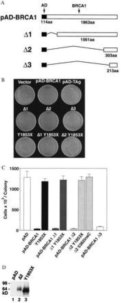
Figure 2.

The C-terminal BRCA1 fragment (fused to the GAL4 activation domain and the nuclear localization signal of simian virus 40 large T antigen) is sufficient for small colony formation. Truncating mutations revert the small colony phenotype. (A) Schematic representation of pAD–BRCA1, pAD–BRCA1–Δ1, pAD–BRCA1–Δ2, and pAD–BRCA1–Δ3. Filled bar indicates GAL4 activation domain fused to the nuclear localization signal of simian virus 40 large T antigen; open bar indicates BRCA1 sequences. Thin connecting lines represent regions deleted. Numbers represent number of amino acids (aa). (B) Photograph of HF7c colonies formed after transformation with the indicated plasmids. Y1853X, pAD–BRCA1 Y1853X; Δ1–Y1853X, pAD–BRCA1–Δ1 modified by Y1853X nonsense mutation, and so forth. (C) Number of cells (× 103) per colony after transformation with the plasmids indicated. Single, transformed colonies were resuspended in water and counted on a hemocytometer. Each bar represents the mean of six cell counts ± SEM. All counts were performed by an individual blinded to the vector used. Δ1–Y1853X, pAD–BRCA1–Δ1 modified by Y1853X nonsense mutation, and so forth. (D) Expression of pAD–BRCA1–Δ2 and pAD–BRCA1Δ2 Y1853X in yeast. Anti-HA Western blot detection of pAD–BRCA1–Δ2 (lane 2) and pAD–BRCA1–Δ2 Y1853X (lane 3) in crude yeast lysate from colonies transformed with corresponding vectors. All AD fusion constructs in this study contain an HA epitope detectable by Western blot.
