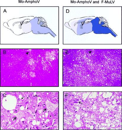
Figure 2.

Neuropathology of DDD mice infected with Mo-AmphoV alone (A–C), or coinfected with Mo-AmphoV and F-MuLV (D–F). (A) Schematic presentation of distribution and severity of spongiform alterations along the neuraxis and hemispheres. Dark-blue shading denotes extreme; medium-blue shading, severe; and light-blue shading, moderate to mild tissue damage. (B) Overview of frontal sections of the medulla oblongata at the level of the foramina of Luschka demonstrating circumscribed foci of spongiform tissue lesions. Ependymum of the fourth ventricle floor is indicated by an arrowhead (×40). (C) Higher magnification (×245) reveals isolated single, large vacuoles of the neuropil, perikaryal swelling, and vacuolization of oligodendrocytes and astrocytes (thin arrows) and two hypertropic, swollen neurons (thick arrows) with pale nuclei and loss of Nissl substance in the cytoplasm. (D–F) Same as A–C, respectively, but mice were coinfected with Mo-AmphoV and F-MuLV. (D) Note lesions aggravate, extend more rostrally, and also involve the hemispheres. (E) In contrast to mice infected with Mo-AmphoV alone, widespread lesioning is observed. (F) Lesions in mice coinfected with F-MuLV are characterized by a higher degree of spongiform degeneration with confluent vacuolization and loss of neurons; the arrows point to shrunken, degenerated neurons.
