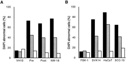
Figure 2.

Apoptin activity in normal versus transformed/malignant cells. The percentage of cells that stained abnormally with DAPI is given as a relative measure for apoptosis in (A) normal (VH10) versus transformed (pre, post) and tumor (NW-18) fibroblasts and (B) normal (FSK-1) versus transformed (SVK14, HaCaT) and tumor (SCC-15) keratinocytes transiently transfected with pCMV-fs (solid bars), pCMV-tr (shaded bars), or pCMV-des (open bars). Cells were fixed 5 days after transfection and analyzed by indirect immunofluorescence. Results are the means of at least three independent experiments. In each experiment at least 200 cells were examined that were positive for full-sized or truncated apoptin, or for desmin.
