
Figure 3.
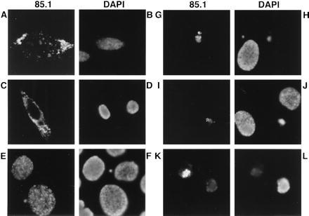
Localization of apoptin and staining of the chromatin in normal and transformed/malignant cells transiently transfected with pCMV-fs. Apoptin was stained with anti-apoptin mAb 85.1 and DNA with DAPI in representations of identical cells: (A and B) VH10, (C and D) FSK-1, (E and F) HaCaT, (G and H) post, (I and J) NW-18, and (K and L) SCC-15. Cells were fixed 2 (E and F) or 5 (A–D and G–L) days after transfection and analyzed by indirect immunofluorescence. (Original magnification: A–D, ×630; I–L, ×1,000; and E–H, ×1,250.)