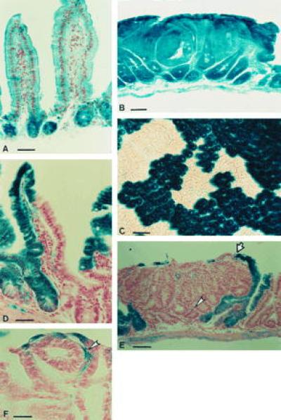
Figure 1.

X-Gal staining in ROSA26 and chimeric mice. After X-Gal staining, samples were post-fixed in 10% neutral buffered formalin, cleared in 70% ethanol, processed, and serially sectioned at 10 μm. Sections were counterstained with Nuclear Fast Red (A, B, and D–F). In R26 mice, staining is observed in all cells of the small intestinal epithelium (A). The β-galactosidase activity is maintained in intestinal adenomas from R26/+ Min/+ mice (B). Crypts in the colon of R26/+ ↔ +/+ mice are monoclonal (C). Villi in the small intestine of R26/+ ↔ +/+ mice are polyclonal (D). Epithelial cells within tumors in R26/+ ↔ Min/+ mice are derived from the Min/+ lineage and are therefore unstained with X-Gal. The normal epithelial layer encapsulating the tumors is polyclonal (indicated by arrows). Scattered X-Gal-positive cells (indicated by arrowheads) in these tumors are lymphocytes and stromal cells (E, F). [Bars = 40 μm (A, B, and E), 80 μm (C), 20 μm (D), and 15 μm (F).]
