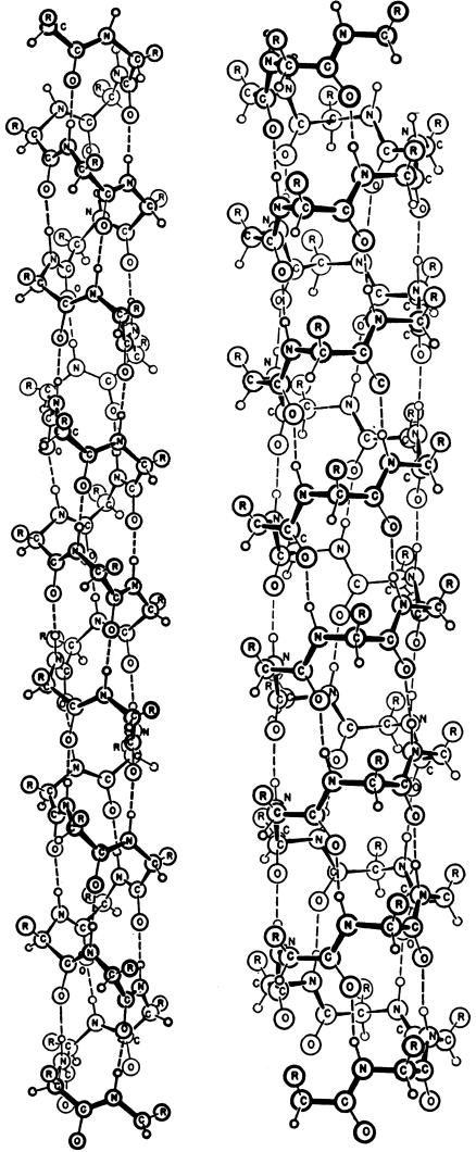
Fig. 2.
The α-helix (Left) and the γ-helix (Right), as depicted in the 1951 paper by Pauling, Corey, and Branson (1). Biochemists will note that the C O groups of the α-helix point in the direction of its C terminus, whereas those of the γ-helix point toward its N terminus, and, further, that the α-helix shown is left-handed and made up of d-amino acids. (Reproduced with permission from Linda Pauling Kamb.)

