
Figure 2.
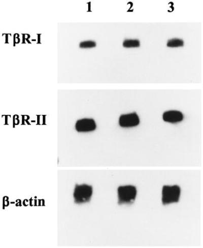
Northern blot analysis of TβRII and TβRI expression in freshly isolated B-CLL cells. Shown are the results from three representative CLL patients. Lanes 1, 2, and 3 correspond to Patients 1, 2, and 3, respectively. The same blot was hybridized to a 32P-labeled probe specific for TβRI, stripped and reprobed using a 32P-labeled probe specific for TβRII, and stripped and reprobed with a 32P-labeled probe to β-actin to verify equal loading of mRNA.