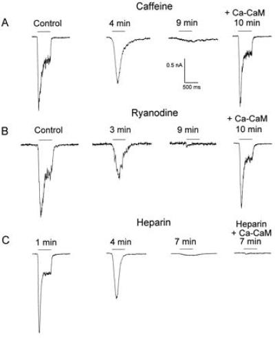
Figure 1.

Chemically induced disruption of the photoresponse in WT cells and its rescue by Ca–CaM. Inward currents are shown in response to orange (OG-590 Schott) light pulses [horizontal bars above the traces (log I = −1, A and C; log I = −2, B] at holding potentials of −60 mV. All the experiments were carried out at normal Ringer’s solution. The time from formation of the whole-cell recordings is indicated. Cells were incubated in a ryanodine- or caffeine-containing solution for 12–40 min. Control photoresponses were recorded before drugs application. Following the disruption of the LIC, internal dialysis with Ca–CaM (5 μM CaM plus 10 μM Ca2+, in other cells of the same retina) rescued the LIC in the ryanodine- or caffeine-treated cells (A and B, fourth trace). Disruption of the LIC was defined as a reduction, to less then 1%, of the geometrical average of the ratios between the peak LIC 10–12 min after drug application and the peak LIC before drug application. A restored LIC was obtained when this ratio was not statistically different from control LIC of 100% (Student’s t test, P > 0.05). The control result was obtain by the same experimental procedure but without application of the chemicals. Throughout the experiments n = 4–7 in each experiment. (A and B) Perfusion of cells with Ringer’s solution containing caffeine (A, 10 mM in 0.1% dimethyl sulfoxide) or ryanodine (B, 4 μM in 0.1% dimethyl sulfoxide). Application of 0.1% dimethyl sulfoxide to the bath for 1 h had virtually no effect on the LIC (n = 4). (C) Heparin sulfate (10 mg/ml) was applied through the whole-cell recording pipette. The first trace shows a normal LIC recorded 1 min after formation of whole-cell recordings, before heparin had any significant effect. Internal dialysis with Ca–CaM (5 μM CaM plus 10 μM Ca2+) together with heparin did not rescue the LIC (fourth trace, recorded from another cell of the same retina).
