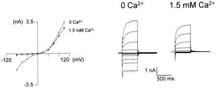
Figure 6.

ISOC arises from activation of the light-sensitive channels. Increasing external [Ca2+] inactivated ISOC in WT photoreceptors. The I–V curves were measured 2–8 min after ISOC was induced by ryanodine plus caffeine (4 μM ryanodine and 10 mM caffeine), and examples of curves from a single cell are presented. The pipette solution included ions that blocked K+ channels. Increasing external Ca2+ inactivated the inward current. Current traces were elicited by 500-ms voltage steps (−80 to +100 mV in intervals of 20 mV) in a cell bathed in Ca2+-free medium (at a holding potential of −20 mV; 0 Ca2+, ○) and 2 min after 1.5 mM Ca2+ was added to the bathing solution (1.5 Ca2+, •, recorded from the same cell). The graphs are the current–voltage relationships (I–V curves) measured from the peak amplitudes of the presented currents (right traces, n = 4).
