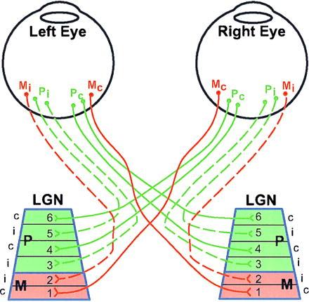
Figure 1.

Schematic illustration of neuronal connections between the eyes and the LGN in the macaque monkey. The connections subserving the left and right eyes might be either parvocellular (P, about 80%), magnocellular (M, about 10%) or others (K, I, or S about 10%, not shown). The different types of ganglion cells in the retina are intermixed, although the percentage of P cells is higher in the central retina than at the periphery. In the LGN, layers 1 and 2 are magnocellular (red), and layers 3–6 parvocellular (green). The set of M ganglion cells in a given eye that project to the contralateral LGN terminate in layer 1, whereas those that project ipsilaterally terminate in layer 2. The P ganglion cells project to layers 3 and 5 to the LGN on the same side, and 4 and 6 on the opposite side. For more details see ref. 1–7.
