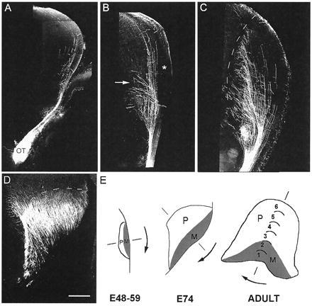
Figure 5.

Photomontages of confocal images through representative coronal sections of the diencephalon showing the distribution of DiI-labeled retinal axons in the embryonic thalamus. (A–D) Dashed lines indicate the border of the dorsal lateral geniculate (dLGN) with a dorsal orientation to the top and a lateral orientation to the right. (A–C) The contralateral side to the DiI optic nerve implants. (A) At E48, retinal axons navigate the contralateral optic tract before being deflected away from the pial surface as they approach the geniculate. A few axons course dorsally past the dLGN toward the midbrain. (B) At E53, there is greater ingrowth of retinal fibers, some of them elaborating medially directed branches. Note that the lateral aspect of the dLGN remains totally devoid of retinal axons (∗). Branches derived from the axon trunks are concentrated at the bottom third of the dLGN, and in some cases these extend past its medial border in the external medullary lamina (arrow). (C) At E64, an increasing number of axonal branches invade the medial region of the nucleus. Note that, at this age, the lateral segment of the dLGN is still virtually free of retinal afferents. The section shown is from the rostral part of the dLGN, but essentially the same pattern is observed throughout the rostro-caudal extent of the nucleus. (D) At E74, DiI crystals were implanted into the optic tract to reduce the distance of diffusion. Virtually the entire extent of the nucleus now receives a retinal innervation. The coronal section is from the caudal aspect of the dLGN. (E) Schematic representation of the dLGN rotation (arrows) from E48 to adulthood. The presumptive M layers (shaded area) rotate from a lateral to a ventral position whereas the presumptive P layers rotate from a medial to a dorsal position (46). (Scale bar: A–C, and E, 500 μm and D, 400 μm.)
