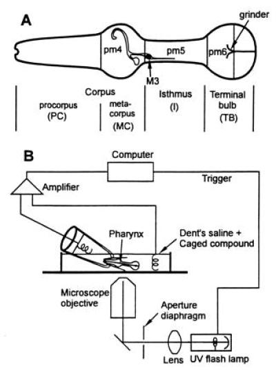
Figure 1.

(A) Anatomy of the C. elegans pharynx (modified from ref. 3). Anterior is to the left. The pharynx is divided into three major functional parts, the corpus in the front of the nematode [subdivided into procorpus (PC) and metacorpus (MC)], I, and TB, and contains 20 neurons and 37 muscle cells (3). The location of the nucleus of the M3 motor neuron on the left side of the pharynx is shown; another M3 motor neuron is in the corresponding location on the right side of the pharynx. The location and extent of three of the large muscle types are indicated: pm4 in the MC, pm5 in the I area, and pm6 in the front region of the TB. The process of the M3 neuron to pm4 and pm5 muscle cells is drawn as an open line in this figure. (B) Diagram of the instrumentation.
