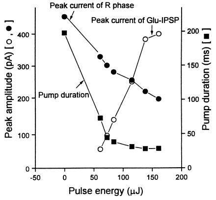
Figure 3.

Data obtained with EPGs from MT6308 (eat-4) mutants that are defective in M3 function and that do not exhibit IPSPs in their EPGs (R. Y. N. Lee, personal communication; ref. 6). By varying the pulse energy (from 61 μJ to 161 μJ), the concentration of glutamate released by photolysis of 0.5-mM caged glutamate in an illuminated area of about 200 μm diameter was increased. The UV flash lamp was turned on 20 ms after the appearance of the E spike in the EPG. The Glu-IPSP increased and the pump duration and the R-phase spike current in the EPG decreased with increasing pulse energy used. On average five experiments were done for each data point shown. The SEM is similar to the size of the symbol in the figure. A similar relationship was obtained by changing the concentration of caged glutamate in the bath (from 0.25 mM to 1.0 mM) but keeping the pulse energy and illuminated area constant (data not shown).
