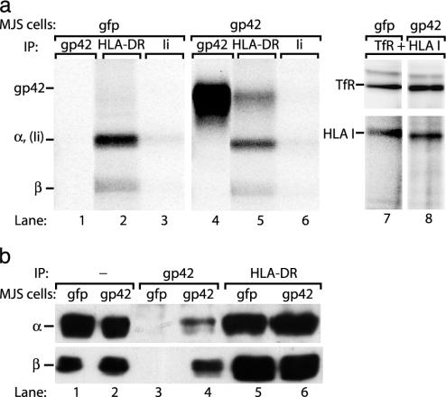
Fig. 1.
Endogenously expressed EBV gp42 associates with HLA class II molecules. (a) Surface-exposed proteins on MJS/gp42 (lanes 4–6 and 8) and control MJS/gfp (lanes 1–3 and 7) cells were labeled with 125I for 1 h. Specific proteins were isolated from cell lysates by immunoprecipitation (IP) with mAbs against gp42 (F-2-1), HLA-DR (Tü36), HLA class I (W6/32), and TfR (66IG10); Ii (VicY1) served as a negative control. Precipitated proteins were separated under nonreducing conditions by SDS/12% PAGE. (b) Total lysates of MJS/gp42 (lanes 2, 4, and 6) and control MJS/gfp (lanes 1, 3, and 5) cells were directly loaded (lanes 1 and 2) or were subjected to immunoprecipitations with rabbit serum no. 32 against gp42 (lanes 3 and 4) and mAb Tü36 against HLA-DR complexes (lanes 5 and 6). Total lysates and immune complexes were boiled in reducing (α) or nonreducing (β) sample buffer, separated by SDS/12% PAGE, and blotted onto poly(vinylidene difluoride) membranes. Western blots were stained with mAbs specific for HLA-DRα (DA6.147) and β (HB10A) chains and visualized by enhanced chemiluminescence.

