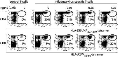
Fig. 4.
TCR interactions with complexes of peptide–HLA class II, but not class I, are abolished in the presence of soluble EBV gp42. Peripheral blood mononuclear cells from a healthy HLA-A2+ DR4+ donor were labeled with CFSE and stimulated in vitro with DR4/HA307–319 (Upper) or A2/M58–66 (Lower) or without peptides (control). Responding T cells were incubated with specific allophycocyanin-conjugated tetramers in the presence or absence of rgp42. rgp42 concentrations of 0.05, 0.25, and 1.25 μM correspond to gp42:class II ratios of 0.5:1, 3:1, and 12:1, respectively. Cells were stained with phycoerythrin-conjugated mAbs to CD4 (Upper) or CD8 (Lower). Each flow-cytometry dot plot represents ≈45,000 propidium iodide–, CFSE– cells; percent values refer to the percentage of CD4+ or CD8+ T cells that stained with the tetramers (top right quadrant).

