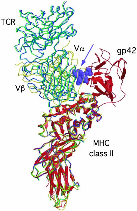
Fig. 5.
EBV gp42 sterically clashes with the TCR in known TCR–MHC class II structures. Superposition of the gp42–DR1 structure (shown as red ribbons) with the crystal structures of the HA1.7-TCR–DR1 complex (green), the HA1.7-TCR–DR4 complex (blue), and the D10-TCR–I-Ak complex (yellow). The gp42 loop including residues 157–161 clashes with the TCR Vα domains and is shown as purple Corey–Pauling–Koltun model atoms (arrow). The D10-TCR has the greatest overlap with gp42, whereas the HA1.7 structures are rotated slightly away from gp42.

