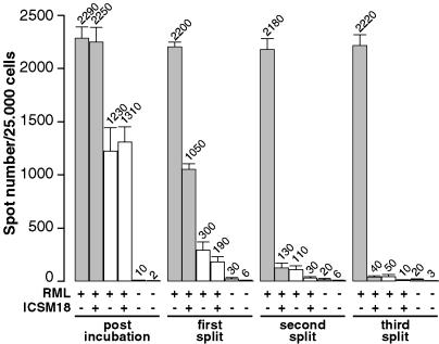
Fig. 2.
Clearance of PrPSc particles from the initial inoculum after three successive 1:10 splits of infected cells. Twenty thousand susceptible N2aPD88 cells (gray bars) and nonsusceptible NN2a cells (white bars) were cultured for 16 h, plated in a 96-well plate, and exposed for 3 days to a 10–4 dilution of RML strain (I2424) in OFCS, in the absence (–) or presence (+) of the anti-PrP mAb ICSM 18 (5 μg/ml, added 4 h before infection), as indicated. As controls, N2aPD88 and NN2a cells were incubated with OFCS only. Three days after infection, the cells were suspended, octaplicate aliquots of 25,000 cells were filtered onto the membranes of an ELISPOT plate, and the PrPSc-positive signals were quantified by the SC assay. Both susceptible and nonsusceptible cells gave rise to PrPSc-positive signals. An anti-PrP antibody that abrogates PrPSc accumulation did not diminish the spot number, demonstrating that all spots can be accounted for by PrPSc particles from the inoculum. After three 1:10 splits, nonsusceptible and susceptible cells incubated with antibody showed <2% of the spots given by susceptible cells. The numbers for noninfected N2aPD88 cells and NN2a cells, the fifth and sixth bar of each group, respectively, represent the background. Mean values and SD are shown.

