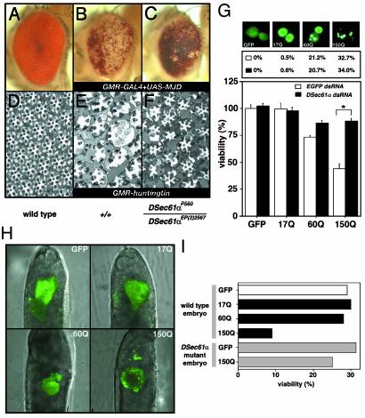
Fig. 3.
The DSec61α translocon is involved in expanded polyglutamineinduced neural cell death and degeneration in Drosophila. (A–F) The degenerative eye phenotype of the different Drosophila models for polyglutamine disease was improved in mutants for the DSec61α gene. Light microscopic (A–C) and semithin section (D–F) images of the compound eyes are shown (day 30 after eclosion). (Magnifications: ×100.) The following genotypes are shown: w;GMR-GAL4/+ (A and D), w;GMR-GAL4/+;UAS-MJD(M)/+ (B), w;GMR-GAL4 DSec61αP560/DSec61αEP(2)2567;UAS-MJD(M)/+ (C), w;+/+;GMR-huntingtin120Q/+ (E), and w;DSec61αP560/DSec61αEP(2)2567;GMR-huntingtin120Q/+ (F). (G) The neural cell death induced by expanded polyglutamine tracts was attenuated in the DSec61α gene knockdown cells. (Top) Plasmids (200 ng of each type) encoding the expanded polyglutamine tracts with the first exon of human Huntingtin fused to EGFP (pUAST vectors) were overexpressed in Drosophila BG2 neural cells with 200 ng of driver plasmid pWAGAL4. The fluorescence indicating GFP-positive cells was examined 24 h after transfection. (Middle) Plasmids (200 ng of each type) encoding the expanded polyglutamine tracts were cotransfected with 25 ng of synthesized dsRNA (EGFP or DSec61α) in Drosophila BG2 neural cells with 200 ng of driver plasmid pWAGAL4. The numbers indicate the percentages of the population of GFP-positive cells that contained fluorescent aggregates (EGFP dsRNA, ○; DSec61α dsRNA, •). (Bottom) Plasmids (5 ng of each type) encoding the expanded polyglutamine tracts were cotransfected with 25 ng of synthesized dsRNA (EGFP or DSec61α) in Drosophila BG2 neural cells with 2 ng of driver plasmid pWAGAL4, and cell viability was determined. (H and I) The aggregate-like structure was produced by expanded polyglutamine in Drosophila embryos, which affected viability of embryos. Three types of expanded polyglutamine tracts with the first exon of human Huntingtin fused to EGFP and 100 ng/μl pCaspeR-hs vectors were injected into Drosophila embryos (w1118 and DSec61αEP(2)2567/DSec61αP560). At 12 h after injection, embryos were heat-shocked at 37°C for 1 h and developed at 25°C for 12 h. The fluorescence indicating GFP-positive cells was examined 18 h after injection (H). And then the number of hatched larvae was counted (I).

