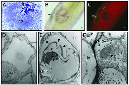
Fig. 1.
Plant cell death during infection pocket formation. (A) Toluidine blue-stained section through a 3-day-old developing root nodule. Evans blue-stained lateral root base, 2 days after inoculation with ORS571(pBBR5-hem-gfp5-S65T) viewed with bright-field optics (B) or epifluorescence (C). TEM of infection pocket region of stem-borne developing nodules 4 days after inoculation: healthy cortex cell (D), early stage of cell death (E) (arrows indicate disruption of wall–plasma membrane integrity), and dead plant cell at the edge of infection pocket (F) (arrow shows invaded bacteria). ic, inner cortex; ip, infection pocket; np, nodule primordium; vf, vacuole fragmentation. [Bars = 100 μm (A–C) and 10 μm (D–F).]

