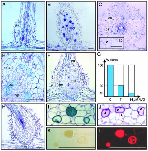
Fig. 3.
Nod factor and ethylene effects on lateral root bases. Roots were treated with 10–9 M Nod factors or ethylene for 5 days. All sections except J–L were stained with toluidine blue. (A) Mock-inoculated root. Arrow indicates intercellular spaces. (B and C) Ethylene treatment. (D) Detail of C; arrowhead indicates cavity with cell wall remainders. (E and F) Nod factor treatment. (G) Percentages of plants that show lateral root base enlargements on Nod factor application after pretreatment with AVG. (H) Example of a Nod factor-induced swelling after pretreatment with AVG. (I) Toluidine blue-stained section through a Nod factor-treated lateral root base. (J) Differential staining of analogous section of I. Dark-blue nuclei (arrows) indicate cells are dying; arrowhead marks nucleus of a healthy cell. (K) Unstained neighboring section of I under bright-field optics. (L) Epifluorescence microscopy of K. For abbreviations, see Fig. 1; bc, blue cell; ca, cavity. (Bars = 100 μm.)

