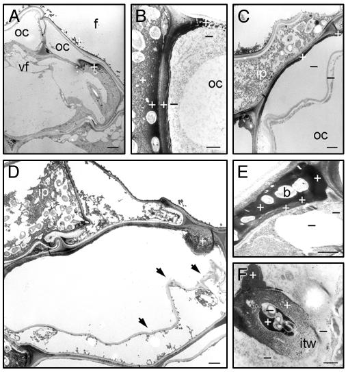
Fig. 4.
H2O2 localization during nodule initiation. TEM sections of CeCl3-stained adventitious rootlets, harvested at 50 (A) and 70 (B–F) hpi with ORS571. Plus or minus indicates presence or absence of Ce perhydroxides, confirmed by electron probe x-ray microanalyses. (A) Dying outer cortex cell. (B) Infection pocket-cortical cell boundary. (C) Outer cortex cell neighboring infection pocket. (D) Papillus-forming dying outer cortex cell; arrows indicate disruption of wall-plasma membrane integrity. (E) Intercellular infection thread. (F) Intracellular infection thread. For abbreviations, see Fig. 1; b, bacteria; cw, cell wall; f, fissure; itw, infection thread wall. (Bars = 1 μm.)

