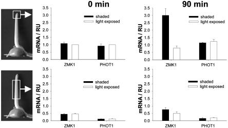
Fig. 3.
Localization of ZMK1 and PHOT1 expression in the photostimulated coleoptile. Relative transcript levels (RU, relative units) of ZMK1 and PHOT1 in light-exposed (open bars) and shaded (solid bars) halves of the coleoptile tip (Upper) and base (Lower) after 0 (Left) and 90 (Right) min of stimulation with unilateral blue light. The mRNA content (n = 3 ± SE) was quantified as described in Materials and Methods. The level of ZMK1 and PHOT1 mRNA in the light-exposed half at t = 0 min was set to 1.0.

