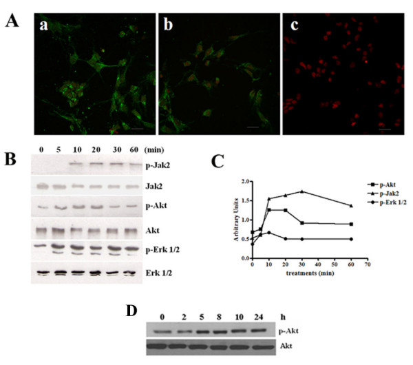
Figure 1.
Expression and activation of IL-3 receptors in cortical neurons. (A). Primary cortical neurons were incubated with anti-IL-3α subunit (a), anti-IL-3β subunit (b) or without first (c) antibodies followed by incubation with a second antibody conjugated to FITC. Scale bar, 20 μm. (B). Western blot analysis shows Jak2, Akt and ERK phosphorylation in cortical neurons treated with 5 nM IL-3 for the indicated times. (C). Normalized densitometry scans of proteins panel B. (D). Western blot analysis shows Akt phosphorylation in cortical neurons treated with IL-3 for the indicated times. The results are representative of three separate experiments.

