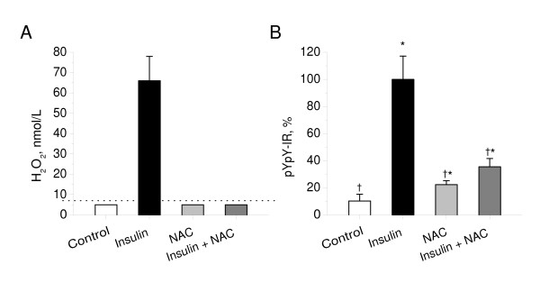
Figure 1.
Effect of N-acetylcysteine on Insulin-stimulated H2O2 production and the insulin receptor autophosphorylation in cerebellar granule neurons. A: CGN cultures were pre-incubated for 30 min in the absence or presence of N-acetylcysteine (5 mmol/l) in Hepes-buffered salt solution and then exposed to insulin (100 nmol/L). H2O2 release from cultures for 1 min was measured as described in Materials and Methods. Results were normalized by cell density. Columns represent the means ± SD of H2O2 values obtained from five to nine cultures. Dotted line represents a detection limit of the assay (7 nmol/L). B: CGN cultures were pre-incubated for 30 min in the absence or presence of N-acetylcysteine (5 mmol/l) in Hepes-buffered salt solution and then exposed to insulin (100 nmol/L) for 20 min. Autophosphorylation of insulin receptor was measured as described in Materials and Methods. In each experiment, amount of phosphorylated insulin receptor β-subunit (pYpY-IR) was normalized to total amount of insulin receptor β-subunit and expressed as a percentage of the response produced to 100 nmol/L insulin. Columns represent the means ± SD of pYpY-IR values obtained from four to nine culture dishes. *P < 0.05 vs. control.†P < 0.05 vs. insulin.

