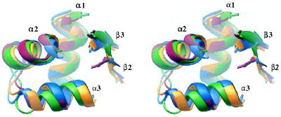
Figure 2.
Stereo view of the superposed backbones of CAP-like regions from E. coli DNA topoisomerase I domain III (red), DNA topoisomerase I domain IV (green), S. cerevisiae DNA topoisomerase II (blue), and E. coli GyrA (yellow). The domains are oriented such the second helix of the HTH motif is facing forward. Figure generated by ribbons (50).

