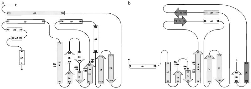
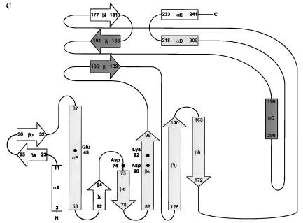
Figure 2.
Schematic representations of the topology of the secondary structure for λ-exonuclease (a), PvuII (b), and EcoRV (c). Regions that structurally correspond in all three proteins are shaded in gray. Regions of structural similarity between PvuII and EcoRV that are not seen in λ-exonuclease are shaded in dark gray. b and c are modified from refs. 15–17. Regions of secondary structure for λ-exonuclease were determined by using the program dssp (29). Structurally corresponding active site residues also are identified.


