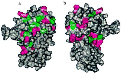
Figure 7.
Surface of the cytohesin-1 Sec7 domain colored by chemical shift changes on binding to Δ17Arf-1: green indicates residues that have chemical shift changes >0.05 ppm in 1H and/or >0.25 ppm in 13C, magenta indicates residues that have chemical shift changes >0.05 ppm in 1H and/or >0.25 ppm in 15N. The orientations are identical to those in Fig. 6. The sites of mutations are annotated.

