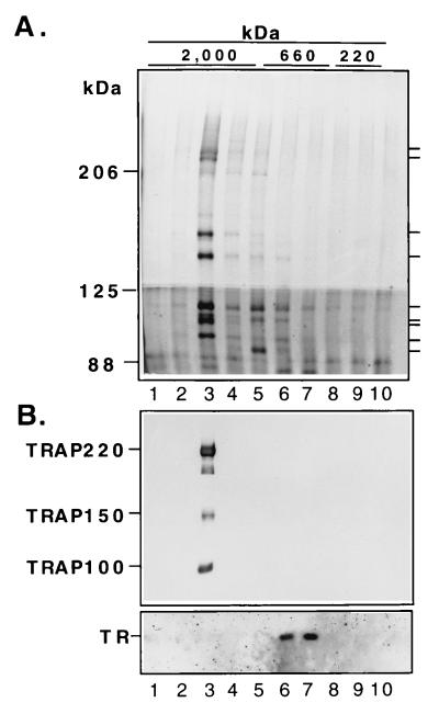
Figure 2.
Gel filtration of purified TR–TRAP complex in the absence of T3. (A) Silver staining. One microgram of TR–TRAP complex was fractionated on Superose 6, resolved by SDS/PAGE with 6% (Upper) and 8% (Lower) acrylamide gels and detected by silver staining. The void volume was not collected. Bars on the right indicate positions of individual TRAPs. (B) Western blot analysis. Proteins (molecular weight <85 kDa) in the lower part of the SDS/PAGE analysis in A were transferred and probed with anti-TRα antibody. Proteins from an analysis equivalent to the SDS/PAGE analysis in A were probed with anti-TRAP220, TRAP150, and TRAP100 antibodies.

