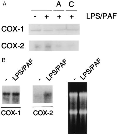
Figure 1.
(A) Expression of COX isoforms in P388D1 cells. The cells were incubated with 5 μM actinomycin D (A) or 10 μM cycloheximide (C) for 30 min before the LPS/PAF treatment (1 h with LPS plus 10 min with PAF). Cells were then lysed as described in the text. Protein (100 μg) was separated by SDS/PAGE and analyzed by immunoblotting using specific antibodies against COX-1 or COX-2. (B) Northern blot of COX-1 and COX-2 from total P388D1 RNA. Total RNA from LPS/PAF-stimulated cells or nonstimulated (−) was isolated and hybridized with probes for COX-1 and COX-2, as indicated. As a control for RNA loading, an “in-gel” RNA picture is shown on the right.

