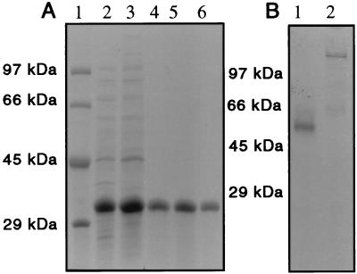
Figure 1.
SDS and native PAGE of the primase fragment. (A) Proteins were separated on a 10% polyacrylamide gel in the presence of 1% SDS. Lanes: 1, 2 μg each of protein standards; 2, 20 μg of Fraction I, a cleared lysate of BL21(DE3) cells containing the plasmid pETg4-PF that were induced with isopropyl β-d-thiogalactopyranoside for 3 hr; 3, 20 μg of Fraction II, the supernatant after the precipitation of nucleic acids with streptomycin sulfate; 4, 5 μg of Fraction III, the DEAE-Sepharose chromatography pool; 5, 5 μg of Fraction IV, the pool from the Sephacryl S200HR column; 6, 5 μg of Fraction V, the pure primase fragment after chromatography on a Hi-trap Blue affinity column. (B) Proteins were analyzed on a native polyacrylamide gel run under nondenaturing conditions. Lanes: 1, 2 μg of primase fragment (Fraction V); 2, 2 μg of the 63-kDa gene 4 protein. The positions of the same protein standards shown in A are indicated.

