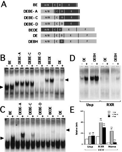
Figure 2.
Comparative gel mobility shift analysis of chimeric receptors in combination with Usp, RXR, or no added heterodimer partner. (A) Schematics of native and chimeric EcRs. BE sequences are depicted with dark shading and DE sequences are shown in light shading. The name of each chimera is to the left of the drawings. Vertical lines represent divides between the different labeled domains characteristic of nuclear hormone receptor family members. (B) Gel mobility shift of native and chimeric EcRs with Usp and vehicle (−) or 1 μM murA (+). The left arrow indicates the size of the native BE shift, and the right arrow indicates the location of the native DE band shift. The lower portions of all of the remaining autoradiograms were essentially identical with A and only the top half is depicted to allow for greater magnification of the area displaying the EcR heterodimer bands in this and subsequent figures. (C) Gel mobility shift of native and chimeric EcRs with RXR as in B. (D) Gel mobility shift of in vitro translated and normalized DE and hinge-substituted DEBH, with Usp or RXR as labeled. −, vehicle; +, 1 μM murA. (E) Transient transfection of VEH into CV-1 cells and directly comparable to Fig. 1B.

