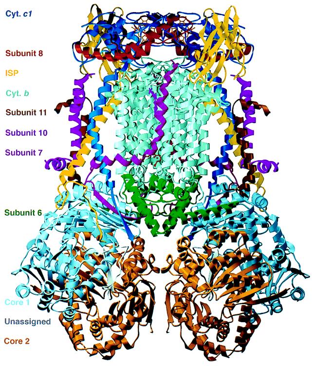
Figure 1.
Ribbon model of the dimeric cytochrome bc1 complex. Colors identifying the subunits are given at the left margin. An as-yet-unassigned peptide is bound in a cavity formed by subunits core 1 and core 2 (16). Figs. 1 and 5 were prepared with the programs molscript (33) and setor (34), respectively.

