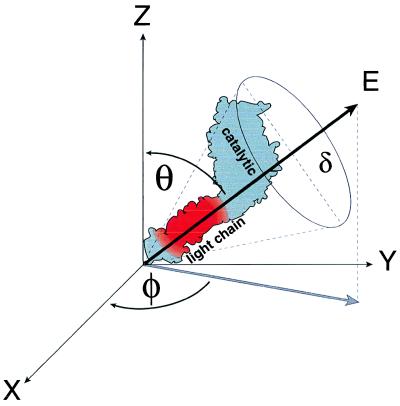
Figure 2.
Spatial model for fluorophore emission dipole. A myosin molecule is illustrated (only the S1 subfragment is depicted with the catalytic and light chain domains labeled), adhered to the plane of the motility surface (x-y plane). The emission dipole electric field vector (E), associated with the 6-IATR-labeled RLC in the myosin light chain domain, is defined in space by its angle, φ, in the x-y plane and angle, θ, relative to the z-axis. The wobble cone for the fluorophore is defined by angle δ, although we have assumed that the fluorophore is rigidly adhered to the RLC.

