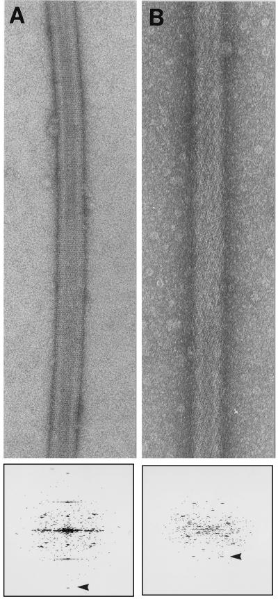
Figure 2.
Helical arrays of two his-tagged proteins on nickel functionalized lipid tubules. (A) Helical array of his tagged Fab 3B3 grown on a nickel functionalized lipid tubule, negatively stained with 1% uranyl acetate. The diffraction pattern below shows visible peaks to 1/19 Å−1 (arrowhead). (Magnification ×140,000.) (B) Helical array of his tagged Fab AP7 grown on a nickel-functionalized lipid tubule, negatively stained with 1% uranyl acetate. The diffraction pattern below shows visible peaks to 1/30 Å−1 (arrowhead). (Magnification ×152,000.)

