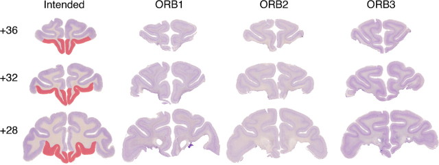
Figure 1.
Lesions of orbital prefrontal cortex. The first column shows the extent of intended damage (red) on sections from the brain of a monkey without damage to orbital prefrontal cortex. The three remaining columns show histological sections from each of the three cases with orbital prefrontal lesions. Each row represents one approximate stereotaxic level, in millimeters anterior to the interaural plane, from anterior to posterior.

