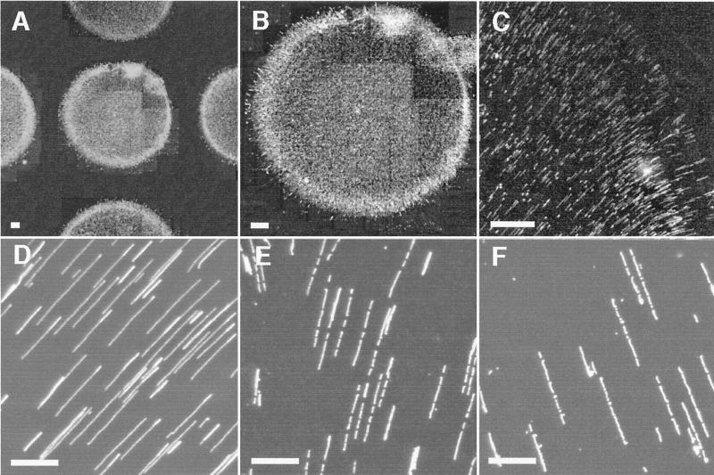
Figure 1.
Digital fluorescence micrographs of gridded spots containing fluid-fixed molecules. Droplets of lambda bacteriophage DNA dissolved in Tris-EDTA buffer containing 0.5% glycerol deposited onto APTES-treated glass surfaces, dried and stained. (A) Section of a 10×10 spot grid on a derivatized surface. Image composed by tiling a series of 16× (objective power) images. (B) Close-up of a DNA spot within the grid. Image composed by tiling a series of 16× images. (C) Elongated DNA molecules on surface before restriction digestion (16×). (D) Magnified image of elongated DNA molecules contained within the spot shown in B before restriction digestion (100×). (E) DNA molecules in B, different field, after digestion with BamHI (100×). Note appearance of gaps signaling enzyme cleavage sites. (F) DNA molecules after digestion with AvaI, from another grid spot, using the same surface and spotting conditions (100×). [Bars: 20 μm (A–C); 5 μm (D–F).]

