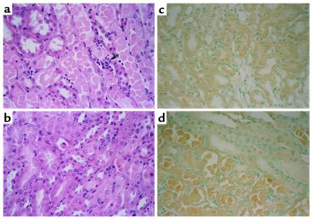
Figure 6.
Renal histopathology (representative picture of at least three experiments). In wild-type mice with ischemic ARF, proximal tubules in the outer medulla show extensive damage including epithelial cell sloughing with focal denudation and numerous neutrophils (arrow) (a). In comparable sections from caspase-1–/– mice, tubules are largely intact with only focal sloughing of tubular cytoplasm and minimal loss of brush border. Neutrophils are inconspicuous in this case (b). Immunohistochemistry for IL-18 showed that there was cytoplasmic immunoreactivity in proximal tubule epithelium in sham-operated wild-type mice (c). There was increased IL-18 staining in necrotic proximal tubule epithelium in ischemic ARF in wild-type mice (d).

