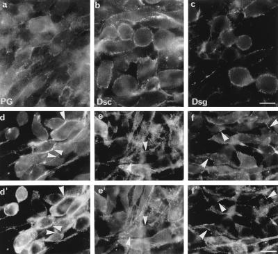
Figure 2.
Immunofluorescent staining of AB5 cells by using antibodies 11E4 (a), HM6 (b), and 3a (c) against PG, Dsca/Dscb, and Dsg1, respectively. (Bar = 18 μm.) Double immunofluorescence of AB5 cells was performed to examine colocalization of PG (d) and Dsg (d′), PG (e) and Dsc (e′), and Dsc (f) and Dsg (f′) by using the antibodies described above. Arrowheads indicate areas of colocalization. (Bar = 12 μm.)

