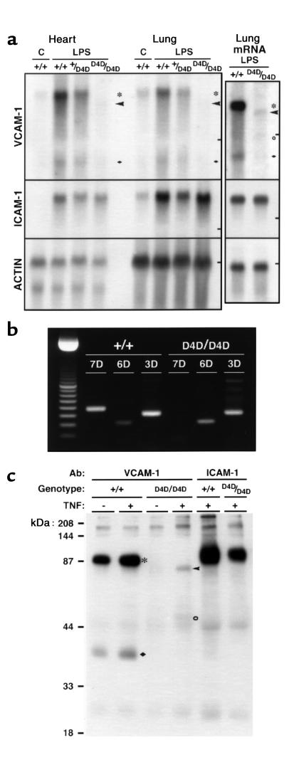
Figure 2.
VCAM-1 expression in Vcam1D4D/D4D mice. (a) Northern blot analysis of VCAM-1, ICAM-1, and β-actin expression using total or oligo dT–enriched RNA (mRNA) from hearts or lungs of control (C) or LPS-treated Vcam1+/+ (+/+), Vcam1+/D4D (+/D4D), and Vcam1D4D/D4D (D4D/D4D) mice. Bars on the right indicate 18S ribosomal RNA migration. Transcripts corresponding to the 7 (asterisks) and 3 (filled diamonds) Ig VCAM-1 were found in Vcam1+/+ and Vcam1+/D4D mice, whereas expression of 6 (arrowheads) and possibly 4 (open circle) Ig domain forms was found in VCAM-1D4D/D4D mice. (b) RT-PCR analysis of 7, 6, and 3 Ig VCAM-1 (7D, 6D, 3D) mRNA in lungs of LPS-treated mice. In Vcam1D4D/D4D mice, domain-specific primers did not detect D4 found in 7 Ig VCAM-1. (c) Immunoprecipitation of cell surface VCAM-1 and ICAM-1 from Vcam1+/+ and Vcam1D4D/D4D biotin-labeled cultured lung endothelial cells. TNF induced expression of 90- to 95-kDa (asterisk) and 36- to 40-kDa (filled diamond) proteins, corresponding to 7 and 3 Ig VCAM-1, in Vcam1+/+ cells, and 80- to 85-kDa (arrowhead) and possibly 50- to 60-kDa (open circle) proteins, corresponding to 6 and 4 Ig VCAM-1 in VCAM-1D4D/D4D cells. No 7 Ig VCAM-1 was detected in VCAM-1D4D/D4D cells. TNF-induced ICAM-1 expression was comparable.

