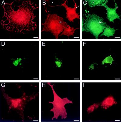
Figure 4.
ClC-5 in transfected cells. COS-7 (A–E, G, and H) or MDCK (F and I) cells were transfected with ClC-5 (A–D, F, G, and I), or, as a control, with ClC-0 (E and H). ClC-5 (detected by PEP5A2) is expressed in vesicles throughout the cytoplasm and in a punctate pattern at the plasma membrane (A and B). Cells in B and C were allowed to endocytose α2-macroglobulin and examined in double-immunofluorescence for ClC-5 (red) (B) and α2-macroglobulin (green) (C). There is a large degree of colocalization as indicated by arrows for some arbitrarily chosen vesicles. A cell not transfected with ClC-5 (∗) has also internalized α2-macroglobulin. Cells in D–I were cotransfected with the GTPase-deficient rab5 mutant Q79L tagged with a myc epitope. Staining for this epitope (green) (D–F) reveals large vesicles representing enlarged early endosomes. These costain for ClC-5 both in COS-7 cells (G) and in MDCK cells that have been grown to an epithelial layer (I). In contrast, when cotransfected with the homologous ClC-0 channel (E and H), ClC-0 is excluded from these vesicles (stained for rab5 in E) and stays in the plasma membrane (as detected with the T12 antibody and shown in red in H). (Bars = 10 μm.)

